
Grey Matter vs White Matter: What’s the Difference?
The brain is often described as if it has two main types of tissue: grey matter and white matter.
That is a useful starting point. It is also one of those tidy explanations that starts to become untidy very quickly if you lean on it too hard.
The simple version is this: grey matter is more involved in processing information, while white matter is more involved in connecting different parts of the nervous system. Grey matter contains many of the brain’s neuron cell bodies, dendrites, synapses, glial cells, and blood vessels. White matter contains many of the long, myelinated nerve fibres that carry signals between regions.
So far, so neat.
But the brain is not a corporate office with a “processing department” upstairs and a “communications department” in the basement. Grey and white matter work together constantly. Grey matter helps generate, interpret, and integrate information. White matter helps that information travel quickly and efficiently across networks.
One without the other would be fairly useless. Grey matter without white matter would be like having lots of busy rooms with no corridors. White matter without grey matter would be an expensive cable system with nobody doing anything at either end.
Which, admittedly, still sounds like some meetings.
What is grey matter?
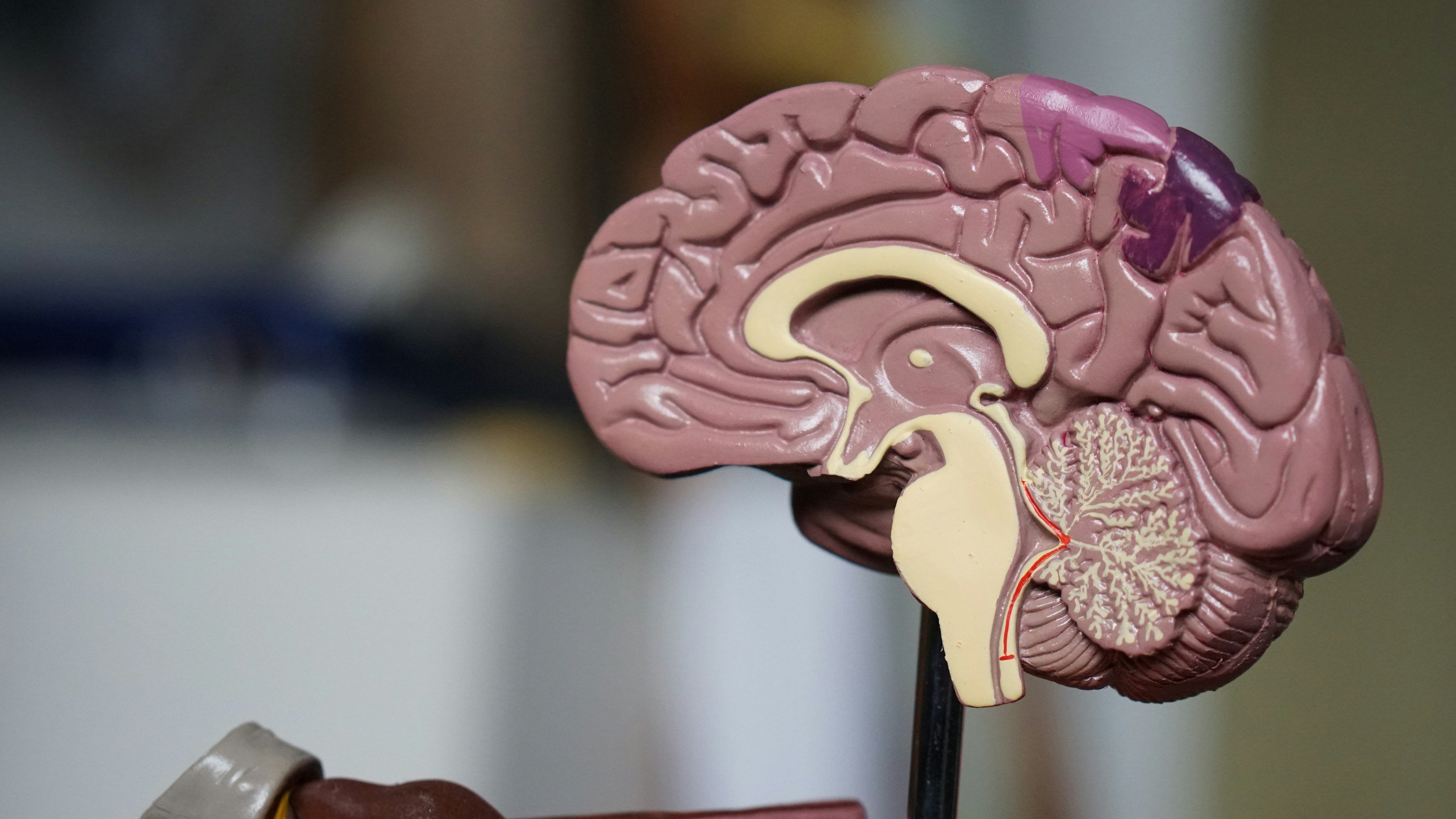
Grey matter is brain and spinal cord tissue that is rich in neuron cell bodies.
Neuron cell bodies contain the nucleus and much of the machinery that keeps a neuron alive and functioning. Grey matter also contains dendrites, synapses, glial cells, capillaries, and some axons. It is called grey matter because, without the fatty white appearance of heavy myelination, it looks greyish in preserved brain tissue.
In the brain, grey matter is found in the cerebral cortex, which is the folded outer layer of the brain. The cortex is involved in perception, movement, language, memory, attention, reasoning, emotion, and decision-making.
Grey matter is also found in deeper brain structures, including the thalamus, basal ganglia, hippocampus, amygdala, hypothalamus, and cerebellum. These regions support functions such as memory, emotion, movement, motivation, sensory relay, coordination, and regulation of bodily processes.
So when people say grey matter is the brain’s “processing” tissue, that is broadly useful. Grey matter is where a great deal of local information processing and integration happens.
But it does not work alone. The brain’s grey matter needs communication routes, and that is where white matter comes in.
What is white matter?
White matter is made mostly of axons.
Axons are long fibres that carry electrical signals away from neuron cell bodies and toward other neurons, muscles, or glands. Many axons in white matter are covered in myelin, a fatty insulating sheath that helps signals travel faster and more efficiently.
Myelin is what gives white matter its lighter appearance.
White matter sits beneath the cerebral cortex and forms major communication pathways across the brain. It allows different regions to share information, coordinate activity, and operate as networks rather than isolated lumps of clever tissue.
A clear example is the corpus callosum, a large white matter tract that connects the left and right hemispheres of the brain. Without this kind of long-range communication, brain regions would struggle to coordinate complex functions.
White matter is not glamorous in the way grey matter often gets to be. Grey matter gets associated with intelligence, thinking, memory, and personality. White matter gets treated like wiring, which sounds less romantic. But communication is not a minor detail. A brain region can be wonderfully specialised, but if it cannot talk to the rest of the system, it becomes less useful very quickly.
The brain is not just built from important places. It is built from connections between them.
Grey matter vs white matter: the simple difference
The easiest way to remember the difference is:
Grey matter helps process and integrate information. White matter helps transmit information between regions.
That is the shortcut.
Grey matter contains more neuron cell bodies, dendrites, synapses, and local circuits. It is heavily involved in analysing sensory input, producing movement, storing and retrieving memories, regulating emotion, supporting language, and making decisions.
White matter contains more myelinated axons. It supports communication between brain areas, between the brain and spinal cord, and between different parts of the nervous system.
But the distinction is not absolute. Grey matter contains axons. White matter contains glial cells and is biologically active, not just passive wiring. Myelin can also change with development, learning, disease, and ageing.
So the better version is this:
Grey matter is where a lot of local neural processing happens. White matter is what helps brain regions work together as coordinated systems.
That less catchy version is also less wrong, which is usually a decent trade.
Where grey and white matter are found
In the brain, grey matter is most visibly found on the outside, in the cerebral cortex. White matter lies underneath it, forming the connecting tracts between cortical and subcortical regions.
There are also important grey matter structures deep inside the brain, including the thalamus, basal ganglia, hippocampus, and amygdala.
In the spinal cord, the arrangement is different. Grey matter sits more centrally, in a butterfly-shaped pattern, while white matter surrounds it on the outside. This arrangement supports the spinal cord’s role in relaying information between the brain and body, as well as coordinating reflexes and movement.
So the old “grey outside, white inside” description works reasonably well for the brain, but not as a universal rule for the whole nervous system. The nervous system, being the nervous system, refuses to be entirely convenient.
What does grey matter do?
Grey matter supports many of the brain’s most complex functions.
Different grey matter regions are involved in different tasks. The visual cortex helps process visual information. The motor cortex helps control voluntary movement. The prefrontal cortex supports planning, inhibition, working memory, decision-making, and social judgement. The hippocampus is important for memory. The amygdala is involved in emotion, threat detection, and emotional learning.
This does not mean each region has one tiny job title and politely sticks to it. Brain functions are distributed across networks. Memory, language, emotion, attention, and decision-making all involve interaction between multiple regions.
Still, grey matter is crucial because it contains many of the local circuits where information is received, processed, compared, and integrated.
If white matter is partly about getting messages around the system, grey matter is where much of the “what does this mean?” work happens.
What does white matter do?
White matter helps the brain communicate with itself.
It supports long-range signalling between regions, allowing different parts of the brain to coordinate. This is essential for attention, learning, memory, movement, language, emotional regulation, and executive function.
For example, reading a sentence involves visual processing, language comprehension, memory, attention, eye movements, prediction, and meaning-making. That is not one brain region heroically doing everything alone while the others make tea. It requires communication across networks.
White matter also helps make neural communication faster and more efficient. Myelin acts like insulation around axons, allowing signals to travel more rapidly. This is especially important for coordinated movement, fast thinking, sensory processing, and complex mental tasks that require different systems to work together.
When white matter is damaged or disrupted, communication between brain regions can become slower, less efficient, or less reliable. This is one reason white matter changes are associated with problems in processing speed, coordination, attention, and executive function.
How grey and white matter develop
Grey and white matter change dramatically across childhood, adolescence, and early adulthood.
Grey matter volume tends to increase during childhood, then decline in some regions during adolescence and early adulthood. This does not mean the brain is simply “losing brain power.” Much of this change is linked to synaptic pruning and refinement. The brain strengthens some connections and reduces others, making networks more efficient.
It is less like a library burning books and more like a very strict editor removing unnecessary footnotes. Still brutal, but with a purpose.
White matter generally increases across childhood and adolescence as myelination continues. As axons become more myelinated, communication between brain regions becomes faster and more efficient. This is one reason cognitive control, processing speed, planning, and coordination often improve across development.
The timeline is not perfectly neat. Different regions mature at different rates, and brain development does not stop on someone’s eighteenth birthday just because paperwork finds that convenient. White matter development can continue into early adulthood, particularly in networks involved in executive function and long-range communication.
Grey matter, white matter, and ageing
Both grey and white matter change with age.
Grey matter volume often declines in later adulthood, although the rate and location of change vary widely. Some regions are more affected than others, and individual differences matter. Genetics, health, activity, education, lifestyle, vascular health, and disease risk can all influence patterns of brain ageing.
White matter also changes with age. Myelin integrity may decline, and white matter lesions or hyperintensities can become more common, especially in relation to vascular health. These changes can affect processing speed, attention, balance, and executive function.
This does not mean ageing equals inevitable collapse. The brain remains adaptable across life, even if it becomes less forgiving of poor sleep, stress, and the kind of admin load no organism should be expected to endure.
A better way to put it is that ageing changes the brain’s structure and connectivity. Those changes can influence cognition and behaviour, but they do not affect everyone in the same way.
Why grey and white matter matter clinically
Grey and white matter differences are important in neurology and psychiatry, but they need to be handled carefully.
In Alzheimer’s disease, grey matter loss is often seen in regions involved in memory and cognition, including the hippocampus and parts of the cortex. These changes are associated with the memory and thinking difficulties that characterise the condition.
In multiple sclerosis, white matter is especially relevant because the condition involves immune-related damage to myelin in the central nervous system. When myelin is damaged, nerve signalling can be disrupted, leading to symptoms such as weakness, sensory changes, visual problems, fatigue, and cognitive difficulties.
In traumatic brain injury, stroke, and other neurological conditions, damage may affect grey matter, white matter, or both depending on the injury and location.
Psychiatric research has also reported grey and white matter differences in conditions such as schizophrenia, major depressive disorder, bipolar disorder, and anxiety disorders. These findings can help researchers understand brain networks involved in emotion, cognition, perception, motivation, and regulation.
But this is where caution is needed.
Brain imaging findings in mental health are often group-level patterns. They do not usually mean that a scan can diagnose depression, schizophrenia, anxiety, or personality from grey and white matter alone. The brain is not a simple barcode for psychological experience, despite everyone’s understandable desire for neat answers and expensive-looking images.
Structural brain differences can be meaningful without being simple.
Common misconceptions
One common misconception is that grey matter is “the thinking part” and white matter is “just wiring.”
This is too crude. Grey matter is heavily involved in processing, but white matter is essential for network communication. Thinking depends on both. If brain regions cannot coordinate, processing suffers.
Another misconception is that more grey matter is always better. It is not that simple. Brain structure needs to be understood by region, age, development, function, and context. More volume in one area is not automatically good, and less is not automatically bad.
A third misconception is that white matter is passive. It is not. White matter changes with development, learning, ageing, and disease. Myelin and axonal integrity play active roles in how efficiently the nervous system works.
Finally, it is worth avoiding the idea that grey and white matter explain personality or intelligence in a simple way. Brain structure contributes to cognition and behaviour, but human psychological life is not reducible to one tissue type behaving itself.
If only.
Simply Put
Grey matter and white matter are two major types of nervous system tissue.
Grey matter is rich in neuron cell bodies, dendrites, synapses, glial cells, and blood vessels. It is heavily involved in processing and integrating information.
White matter is made mostly of myelinated axons. It helps different parts of the brain and nervous system communicate quickly and efficiently.
The simple version is that grey matter processes and white matter connects. That is useful, but incomplete. The brain depends on both working together as part of wider neural networks.
Both grey and white matter change across development, ageing, learning, and disease. They are also relevant in neurological and psychiatric research, but brain imaging findings need care. A difference in grey or white matter is not a simple label, diagnosis, or personality explanation.
In plain terms: grey matter helps the brain do things, and white matter helps the brain do things together.
Which is fairly important, given that the brain is not at its best when its departments stop talking.
References
Braak, H., & Braak, E. (1991). Neuropathological stageing of Alzheimer-related changes. Acta Neuropathologica, 82(4), 239–259. https://doi.org/10.1007/BF00308809
Compston, A., & Coles, A. (2008). Multiple sclerosis. The Lancet, 372(9648), 1502–1517. https://doi.org/10.1016/S0140-6736(08)61620-7
Fields, R. D. (2008). White matter in learning, cognition and psychiatric disorders. Trends in Neurosciences, 31(7), 361–370. https://doi.org/10.1016/j.tins.2008.04.001
Giedd, J. N., Blumenthal, J., Jeffries, N. O., Castellanos, F. X., Liu, H., Zijdenbos, A., Paus, T., Evans, A. C., & Rapoport, J. L. (1999). Brain development during childhood and adolescence: A longitudinal MRI study. Nature Neuroscience, 2(10), 861–863. https://doi.org/10.1038/13158
Lebel, C., & Beaulieu, C. (2011). Longitudinal development of human brain wiring continues from childhood into adulthood. The Journal of Neuroscience, 31(30), 10937–10947. https://doi.org/10.1523/JNEUROSCI.5302-10.2011
Peters, A. (2002). The effects of normal aging on myelin and nerve fibers: A review. Journal of Neurocytology, 31(8–9), 581–593. https://doi.org/10.1023/A:1025731309829
Raz, N., Ghisletta, P., Rodrigue, K. M., Kennedy, K. M., & Lindenberger, U. (2010). Trajectories of brain aging in middle-aged and older adults: Regional and individual differences. NeuroImage, 51(2), 501–511. https://doi.org/10.1016/j.neuroimage.2010.03.020
Wright, I. C., Rabe-Hesketh, S., Woodruff, P. W. R., David, A. S., Murray, R. M., & Bullmore, E. T. (2000). Meta-analysis of regional brain volumes in schizophrenia. American Journal of Psychiatry, 157(1), 16–25. https://doi.org/10.1176/ajp.157.1.16
Table of Contents


Learn about action potential in psychology, a fundamental process in neural communication affecting perception, learning, memory, and behavior. Discover its phases, importance, and link to disorders.